Original Editors – Raquel Lowe
Major Contributors – kim jackson, Raquel Lowe, lucinda hampton, Laura Richie, George Pruden, Joao Costa, Wendy Snyders, Administración, Cheryl Rentchler, Mandé Jooste, Tony Lowe, Evan Tomás, Nikhil Benhur Abburi and Abdallah Ahmed Mohamed
Contents
- 1. Introduction
- 2 Bone Structure
- 3 Joints of the Wrist and Hand
- 4 Ligaments of the Wrist and Hand
- 5 Movements of the Wrist and Hand
- 6 Gripping
- 7 Hand Arches
- 8 Conditions
- 9 Procedures
- 10 References
Introduction
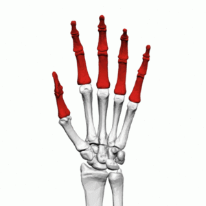
The human hand has a complex mechanism to perform functional capacities. Its integrity is essential for daily functions. Understanding the normal characteristics of the hand requires a thorough analysis of its sensory and mechanical characteristics.(1)
It’s worth watching this 11-minute video.
- The upper limb has sacrificed locomotor function and stability in favor of mobility, dexterity, and precision.
- The hand, located at the end of the upper limb, is a combination of complex joints whose function is to manipulate, grasp, and grip, all made possible by the opposing movement of the thumb.
- Some biologists believe that the development of the human hand indirectly led to the development of our large and complex brain. The existence of the hand promoted brain development by enabling humans to manipulate, interact, explore, and gather information from the environment. A more complex brain, in turn, allowed us to create and use tools, develop a language leading to an elaborate system of shared meanings, known as culture.
Bone Structure
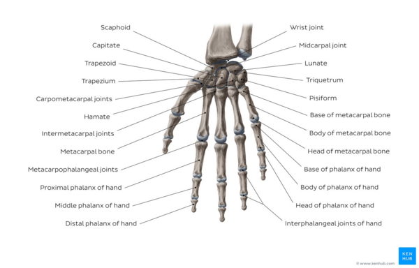
The hand and wrist have a total of 27 bones arranged to roll, rotate, and glide(5); allowing the hand to explore and control the environment and objects. The carpus is made up of eight small bones collectively called carpal bones. The carpal bones are grouped into two sets of four bones:
- pisiform, pyramidal, semilunar, and scaphoid at the upper end of the wrist
- hooked, capitate, trapezoid, and trapezium at the bottom of the hand.
Other hand bones include:
- metacarpals: the five bones that make up the middle part of the hand
- phalanges (singular phalanx): the 14 slender bones that form the fingers of each hand. Each finger has three phalanges (distal, middle, and proximal); the thumb has two.
The hand is divided into three regions.(7)
- The proximal region of the hand is the carpus (wrist).
- The middle region is the metacarpus (palm)
- The distal region is the phalanges (fingers).
Image: Overview of the bones of the wrist and hand.(7)
The Carpus
- The carpus controls the length-tension relationships in the multiarticular muscles of the hand and allows fine grip adjustment.(8)
- Three of the bones in the proximal row articulate with the radius forming the radiocarpal joint and distally with the distal carpal forming the midcarpal joint.
- The four carpal bones in the distal row articulate with the bases of the five metacarpal bones forming the carpometacarpal joints.(9)
- The joints formed between the carpal bones are known as intercarpal joints and most are of the plane synovial type.(3) as the bones interlock with each other, the rows are sometimes referred to as two unique synovial joints.(3)
The arrangement of bones and ligaments allows very little movement between bones.(3)but they slide contributing to finer wrist movements.(10).
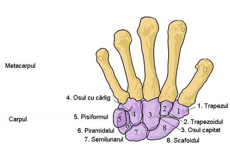
An exception to this is the capitate bone, which has a greater range of motion.(3).
Proximal Row(9)
- Scaphoid – (boat-shaped) – palpable tubercle on the anterior surface. It articulates proximally with the radius, medially with the semilunar, and distally with the head of the large bone. It is a common site of fracture: 70% of all carpal fractures.(9)often injured by a fall on an extended limb
- Lunate – (moon-shaped) – Its palmar surface is smooth and convex and larger than its dorsal surface. Proximally, it articulates with the radius and articular disc, medially with the pyramidal, laterally with the scaphoid, and distally with the head of the large bone.
- Triquetrum – (three-cornered) – Located in the space between the semilunar and the hooked. When the hand is in adduction, it enters the radiocarpal joint.
- Pisiform – (pea-shaped) – a small round bone found in the palmaris longus tendon. It articulates with the palmar surface of the pyramidal. The anterior surface projects distally and laterally forming the medial part of the carpal tunnel.
Distal Row(9)
- Trapezium – four-sided figures with no two parallel sides – most irregular, with a palpable tubercle and an anterior groove medially. It articulates proximally with the scaphoid and medially with the trapezoid. Its articular surface is saddle-shaped and contributes to the mobility of the carpometacarpal joint of the thumb.
- Trapezoid – four-sided figure with two parallel sides – Distally articulates with the second metacarpal, laterally with the trapezium, proximally with the scaphoid, and medially with the large bone.
- Capitate – Head-shaped – the largest of all carpal bones, centrally located and articulated with the semilunar and scaphoid, medially with the hooked, and laterally with the trapezoid. The distal surface primarily articulates with the base of the third metacarpal but also through narrow surfaces with the bases of the second and fourth metacarpals.
- Hamate – hook-shaped – Wedge-shaped with a palpable curved hook projecting from the palmar surface near the base of the fifth metacarpal.
The following mnemonic makes it easier to remember the position of each bone, naming in a circle the carpal bones, starting from the proximal row from the scaphoid to the pinky (little finger) and then the distal row starting from the hooked bone to the thumb:
- See you later for Pinky, here comes the thumb
- Dead straight to Pinky, here comes the thumb
The carpal tunnel – formed by the concave anterior space formed by the pisiform and hooked – on the ulnar side and the scaphoid and trapezium – on the radial side, with a roof-shaped cover of the flexor retinaculum (strong fibrous bands of connective tissue). The long flexor tendons of the fingers and thumb and the median nerve pass through the carpal tunnel
The Metacarpus
The metacarpus, the palm of the hand, composed of five bones: the metacarpals. The bones are numbered laterally, from the thumb, 1 to 5. Each bone is long with a proximal quadrilateral base, a shaft (body), and a rounded distal head. The base of the first metacarpal is saddle-shaped and articulates with the trapezium. The base of the second metacarpal articulates with the trapezium, trapezoid, and large bone. The base of the third metacarpal articulates with the large bone. The bases of the fourth and fifth metacarpal articulate with the hooked bone. The bases of the second through fifth metacarpals also articulate with each other.
The heads of the metacarpals, commonly known as knuckles, are smooth and rounded, extending to the palmar surface; they become

